Traumatic Extensor Hood Rupture
Introduction
Extensor hood rupture is a rare injury associated with boxing and other professional sports.
Extensor tendon rupture occurs secondary to trauma, inflammatory arthropathies (Rheumatoid arthritis) and steroid injection. In boxing, blunt trauma is usually the cause.
Anatomy
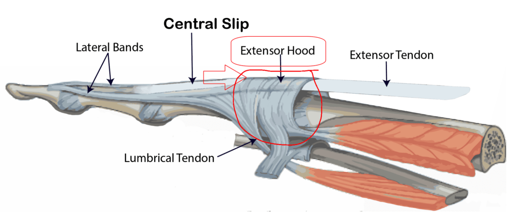
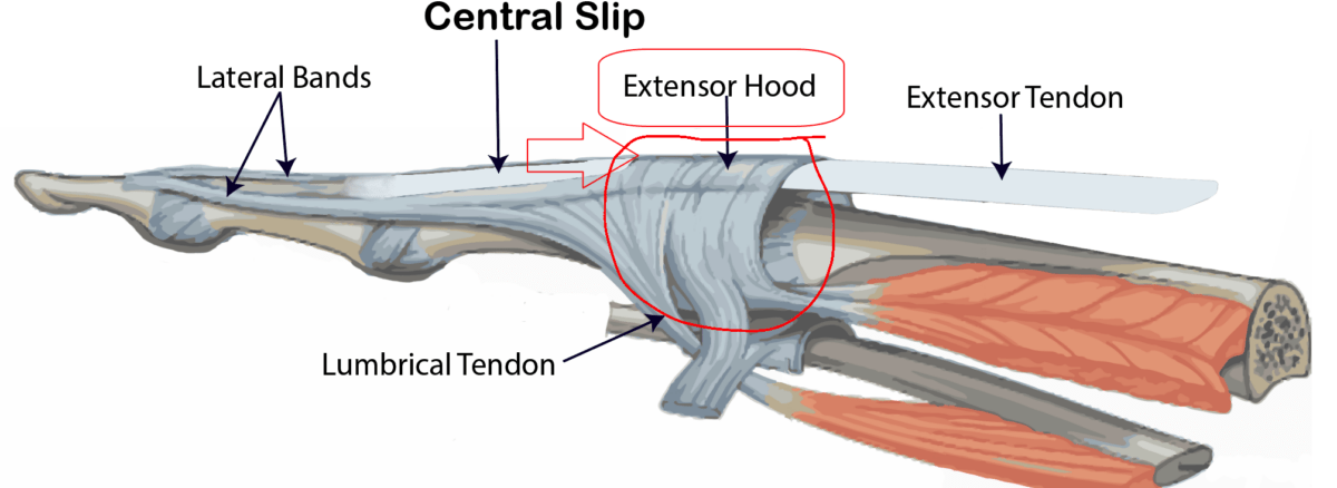
As the extensor tendons travel over the metacarpophalangeal joint (MCPJ), a dorsal sling of transverse fibres is present.
The sling attaches to the extensor tendon dorsally and passes the volar on each side of the MCPJ joint to connect to the volar plate and the transverse metacarpal ligament. This dorsal sling is called the sagittal band. These transverse fibres are layered in a crisscross pattern that changes its arrangement as the finger flexes and extends.
MCPJ joint extension is accomplished by the contraction of the finger extensor tendon and its attachment to the proximal phalanx via sagittal band fibres. The sagittal band acts both as a static tether to prevent displacement of the extensor tendon and as a dynamic tether that allows proximal and distal gliding of the extensor’s tendons during flexion and extension.
Complete laceration of the sagittal bands on either side can cause instability in the extensor tendon position as it travels across the MCPJ joint. Cut to the radial side of the sagittal band causes far more flux than the injury ulnar side. A complete tear of one side of the sagittal band causes subluxation of the extensor tendon to the opposite side of the cut when the MCPJ joint is flexed. When the MCPJ joint is extended, and tension is removed from the Extensor Digiti Minimi (EDM), the tendon snaps back into the central position.
Clinical Picture/Diagnosis
Local tenderness and pain are the first symptoms of the injury.
The pain may limit function, but the patient may be able to carry on for some time. The clinical assessment would reveal a swollen finger and subluxation of the extensor tendon.
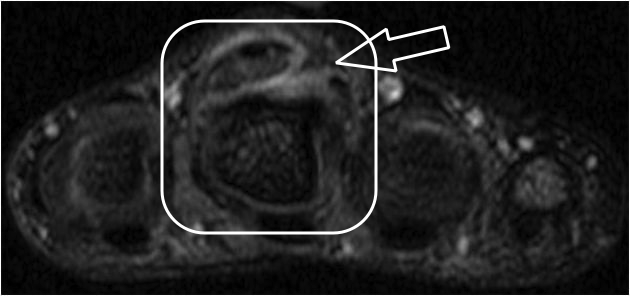
One study utilised magnetic resonance imaging (MRI) to diagnose a ruptured Extensor Hood of the middle finger. It revealed a longitudinal split in the ulnar extensor hood and the joint capsule with marked extravasation of contrast.
Willekens used ultrasonography in 2 patients with a traumatic injury of the index. Patients underwent a second US evaluation one year following the initial trauma. The injuries of the sagittal bands were seen as a hypoechoic thickening at the side of the extensor tendons. Dislocation of the extensor tendons could be observed. The ultrasound examination one year after the trauma revealed an instability of the extensor tendons.
Management
Surgical Intervention
Several studies suggested favourable outcomes associated with surgical repair compared to non-operative treatment.
Loosemore & colleagues conducted surgical repair of the extensor hood with (MCPJ) in 90° of flexion and immobilisation of the MCPJ in flexion for four weeks and reported successful outcomes. The repair was performed with the MCPJ in 90° flexion to avoid stiffness in the Extension. The Edinburgh position was utilised for immobilisation. For four weeks, the MCPJ was placed in 90° flexion with the proximal IP joints in full extension. This position avoids potential shortening, stiffness, and contracture.
Rehabilitation and Functional outcomes
There isn’t much literature regarding rehabilitation after this condition, as for the rare incidence rates. The available evidence concerning surgical restoration discussed the phases after surgery and related them to most hand repair surgeries with similar outcomes, with boxing-specific functions considered.
Normal Anatomy of Extensor hood

MRI scan showing rupture of the extensor hood


