Sesamoiditis
Sesamoid bones are usually present within the substance of the tendon bending the thumb. Sesamoids play important role in the function of the thumb by absorbing pressure, reducing friction at the head of the bone underneath, protecting tendons, and provide a fulcrum for tendons and increasing flexion power. The sesamoid function is analogous to the patella (knee cap) as they increase the mechanical advantage of the flexor tendon of the thumb. The two sesamoid bones of the thumb’s metacarpophalangeal (MCP) joint are embedded in the volar plate. The sesamoid bones articulate with the front aspect of the head of the first metacarpal. A fibrous sheath bridges the front of the sesamoid bones, forming the first annular (A1) pulley floor.
The tendon of the transverse head of the adductor pollicis tendon inserts onto the larger ulnar sesamoid. The smaller, radial sesamoid provides an insertion point for the tendon of the deep head of the flexor pollicis Brevis. The sesamoid bones thus provide a strong point for tendon insertion. This elevates the tendons away from the axis of joint motion, increasing their moment arms and mechanical advantage. The sesamoids can be subjected to trauma or degenerative changes resulting from abnormal tracking on the metacarpal head. This can result in cartilage wear of the articular facets that may manifest as chronic dysfunction and pain—a condition known as chronic sesamoiditis.
Sesamoid provocation test
The thumb MCP joint is stabilised by the thenar intrinsic muscles. When the thumb interphalangeal (IP) joint is flexed against resistance with the MCP joint in flexion (or in a neutral position), the thenar intrinsic muscles contract to stabilise the MCP joint, this elevates the sesamoid bones away from the metacarpal head, increasing the moment arm and mechanical advantage of the thenar intrinsic muscles. In addition, the tendon of the flexor pollicis longus falls palmarly, away from the metacarpal head. However, when the same manoeuvre is done with the MCP joint in hyperextension, the thenar muscles are pulled out to length and cannot lift the sesamoid away from the metacarpal head. The taut flexor pollicis longus tendon presses against the sesamoids, bringing them closer to the metacarpal head.\
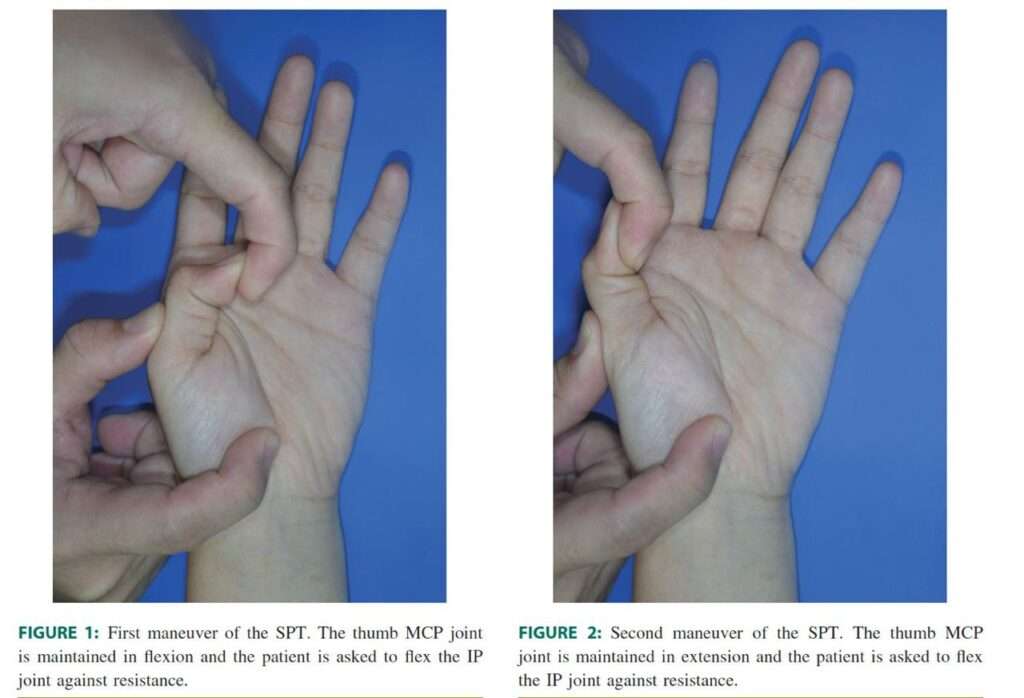
This forms the basis for the SPT. The test consists of two manoeuvres. In the first manoeuvre, we hold the patient’s affected thumb maintaining the MCP joint in flexion and ask the patient to flex the IP joint against resistance (Fig. 1). In the second manoeuvre, we retain the thumb MCP joint in hyperextension, and ask the patient to flex the IP joint against resistance (Fig. 2). The SPT is positive when the patient experiences little or no pain with the first manoeuvreand considerably more pain with the second manoeuvre. The thumb MCP joint is stabilisedby the thenar intrinsic muscles.
When the thumb interphalangeal (IP) joint is flexed against resistance with the MCP joint in flexion (or in neutral), the thenar intrinsic muscles contract to stabilize the MCP joint. This elevates the sesamoid bones away from the metacarpal head, increasing the moment arm and mechanical advantage of the thenar intrinsic muscles. In addition, the tendon of the flexor pollicis longus falls palmarly, away from themetacarpal head. However, when the same manoeuvre is done with the MCP joint in hyperextension, the thenar muscles are pulled out to length and cannot lift the sesamoid away from the metacarpal head. The taut flexor pollicis longus tendon presses against the sesamoids, bringing them closer to the metacarpal head.
This forms the basis for the SPT. The test consists of two manoeuvres. In the first manoeuvre, we hold the patient’s affected thumb maintaining the MCP joint in flexion and ask the patient to flex the IP joint against resistance (Fig. 1). In the second manoeuvre, we retain the thumb MCP joint in hyperextension, and ask the patient to flex the IP joint against resistance (Fig. 2). The SPT is positive when the patient experiences little or no pain with the first manoeuvreand considerably more pain with the second manoeuvre.

Treatment
Treatment modalities have included splints, analgesics, steroid injections, physical therapy, and sesamoidectomy.


