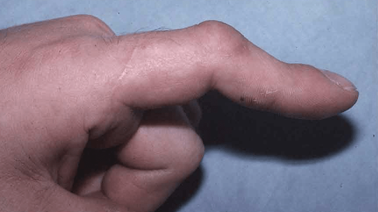
Posttraumatic Finger Contractures and stiffness
Stiffness in the metacarpophalangeal (MCP) joint and proximal interphalangeal (PIP) of the fingers may develop directly due to injury to the joints and adjacent tissues or indirectly due to excessive immobilization or poor splinting of the hand.
Which structures could be involved?
⦁ The circumstances precipitating the contracture determine the structures most involved:
⦁ The joint capsule and collateral ligament contracture
⦁ Flexor tendon adhesions
⦁ Intrinsic musculature contracture
⦁ Extensor tendon adhesions
⦁ Skin and subcutaneous tissue scarring
⦁ The MCP joint (Joint between your hand and your finger) generally stiffen in the straight position.
⦁ The PIP (Joints in your finger) joint often becomes contracted in the flexed position, although extension and combined contractures are not uncommon.
⦁ The key to successfully mobilizing a stiff MCP or PIP joint is anticipating the pathologic causes before surgery.
What is the natural history of the problem?
Long-standing scarring and contracture of the fingers capsule invariably lead to adhesions to the adjacent flexor sheath and extensor mechanism.
Cartilage gradually atrophies and softens with disuse.
Surface irregularities may develop.
How can we diagnose the problem?
Clinical assessment
⦁ History
⦁ The inciting cause of the joint contracture
⦁ The time of the injury
⦁ Efforts made to mobilize the digit
⦁ Assess hand for swelling and the skin status.
⦁ Ongoing swelling and inflammation must subside before surgery if surgery is indicated.
⦁ Prof Imam will assess the active and passive ROM
⦁ Assess the status of the other muscles.
Imaging
Plain radiographs of the hand are made to evaluate for extrinsic and intrinsic causes of joint stiffness.
There is little role for the digits’ computed tomography (CT) scanning or magnetic resonance imaging (MRI).
Other causes of stiffness
⦁ Muscle spasticity or intrinsic muscle paralysis or deficient nerve supply
⦁ Fracture malunion
⦁ Inflammatory disease
⦁ Skin contracture
⦁ Dupuytren disease
What is the best treatment option?
Nonoperative Management
⦁ Nonoperative efforts to improve joint motion must be tried until movement has plateaued and the soft tissues are quiescent.
⦁ As a general rule, inflammation and oedema will subside, and range of motion will improve for a minimum of 3 to 4 months after a traumatic or surgical insult to the hand.
Operative Treatment
The operative treatment involves releasing the constricting soft tissues either using single or double incisions, mainly if the stiffness is not associated with arthritis or persistent subluxation of the joint.
The literature does not provide any specifics as to when to recommend surgery. We usually make this decision when a “functional arc of motion” has not been achieved after a minimum of 3 months of therapy.
There is no absolute functional arc of motion for the joint. In the absence of interphalangeal contractures, we have found that index, middle, ring, and small finger MCP flexion of 30, 35, 40, and 45 degrees, respectively, is generally satisfactory.
Similarly, 45 degrees or more of total PIP motion is usually satisfactory. Flexion contractures greater than 45 degrees are poorly tolerated and may benefit from surgical release.
Extreme flexion contractures (>60 or 70 degrees) may be best managed with joint fusion. The results of a capsulectomy are often disappointing.
Extension contractures are better tolerated, especially if there is flexion to at least 75 degrees.
When a patient has exhausted nonoperative management options, and joint stiffness exceeds the preceding guidelines, surgery for contracture release is considered.


