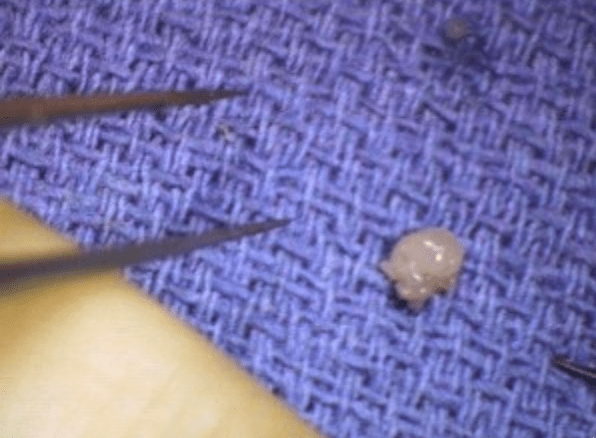
Glomus Tumor
A Glomus Tumour is a rare, mostly benign, soft tissue neoplasm.
The glomus apparatus (or glomus body) is a part of the dermal layer of the skin and is thought to aid in temperature regulation.
When exposed to cold temperatures, the glomus body moves blood away from the surface of the skin to reduce heat loss. While they are located all over the body, glomus apparatus is found in higher concentrations in the fingers and toes. Abnormal growth of a glomus body results in Glomus Tumours.
Glomus Tumors usually occur in people 20 to 50 years of age but are more frequent in young adults. More common in women, 70% of Glomus Tumors present in the hand with the clear majority occurring underneath the nail bed. Most of the nodules are solitary but can occur in clusters. Glomus Tumors represent 1 to 5% of all soft tissue tumors in the hand and fingers.
- 75% occur in the hand
- 50% are subungual
- 50% have erosions of the distal phalanx (primary involvement of bone being very rare)
- less common locations: palm, wrist, forearm, foot
- the glomus body is a perivascular temperature regulating structure frequently located at the tip of a digit or beneath the nail
What causes Glomus Tumors?
Clinical symptoms and diagnosis
Glomus Tumors usually present as a small, firm, reddish-blue bump underneath the finger nail.
These lesions are usually quite small, less than 7mm in diameter.
They can be extremely painful, are sensitive to temperature change, and tender on palpation.
The pain is often worse at night and can be relieved by applying a tourniquet. The mass can cause the nail bed to grow irregularly with ridging possible.
Diagnosis
Glomus Tumours often require a specialist for accurate diagnosis. Upon examination, the mass may appear as a bluish lesion under the nail or in the fingertip pulp. There may be an abnormal ridge in the nail, swelling at the tip and the nodule will be tender to touch. X-rays may show deformity or erosion in the distal phalanx if the mass is long-standing, otherwise, the films may appear normal. MRI (magnetic resonance imaging) is the gold standard for diagnosis.
- pain
- exquisite tenderness to touch
- cold intolerance
- small bluish nodule
- often difficult to see, especially in the subungual location
- nail ridging or discoloration is common
- Love test
- pressure to the area with a pinhead elicits exquisite pain
- 100% sensitive, 78% accurate
- Hildreth test
- tourniquet inflation reduces pain/tenderness and abolishes tenderness to the Love test
- 92% sensitive, 91% specific
Imaging and Tests
X-rays
- glomus tumours can produce a pressure erosion of the underlying bone and an associated deformity of the bone cortex
MRI
- helpful to establish a diagnosis
- Most sensitive Imaging modality
Tests
Treatment
Surgical excision of the tumor is the only treatment modality. During a 15-minute outpatient procedure the nail is removed, an incision is made into the nail bed exposing the tumor, and the tumor is removed. The site is sutured closed and bandaged. The symptoms of pain and cold intolerance are immediately relieved, and the nail will grow back to normal appearance in 3 to 4 months.
Complications
Recurrence : 1 in 5 patients might have a recurrence as per evidence
Summary
- Glomus Tumors are rare benign tumours of the glomus body, often occurring in the subungual region.
- The condition is typically seen in patients between the ages of 20 and 40 who present with a painful subungual mass with bluish discoloration.
- Diagnosis is made with a biopsy showing a well-defined lesion lacking cellular atypia or mitotic activity with the presence of small round cells with dark nuclei.
- Treatment is usually marginal excision.


