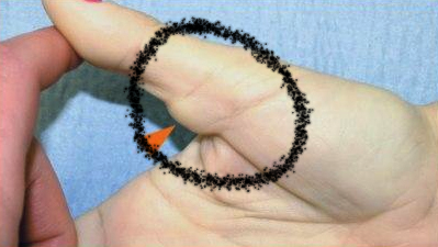
Giant Cell Tumour of Tendon Sheath
It is a common finding which is usually benign.
Second most common soft-tissue tumour seen in the hand, following ganglion cyst
Chassaignac first described these benign soft-tissue masses in 1852, and he overstated their biologic potential in referring to them as cancers of the tendon sheath.
Common in patients aged 30-50 years old
Anatomic location
- is most common on the palmar surface of the thumb, index and middle fingers.
Presentation
- Symptoms and signs: Enlarging mass pain, worse with activity (or wearing shoes, for foot lesions)firm, a nodular mass that does not transilluminate
The symptoms of these masses depend on which subtype you have. Some possible symptoms include the following:
- Swelling that may be painless at first
- Warmth or tenderness around your joint
- Locking, popping or catching when you move your affected joint
- Unstable joint
Diffuse-type giant cell tumors (pigmented villonodular synovitis) can cause degeneration of your joints. It can also damage the bone and cartilage that surrounds your affected joint. If left untreated, it can cause chronic pain and deterioration of your joints.
- Imaging
- Plain radiographs
- To help show the status of the bone underneath
- Ultrasound
- able to demonstrate the relationship of the lesion with adjacent tendon
- most have some internal vascularity
- MRI
- MRI may be helpful diagnostically
- Plain radiographs
- Other causes to consider?
- Ganglion cyst
- cystic component
- Pigmented villonodular synovitis (PVNS)
- histologically identical
- involves larger joints
- Desmoid tumour
- fibroma/fibrosarcoma
- glomangioma
- Ganglion cyst
Treatment
Surgery. Surgery is the main treatment for tenosynovial giant cell tumours. Prof Imam may remove some or all of the tumours, as well as the inflamed joint tissue. You may need another surgery if the tumour returns.
Surgical procedure: marginal excision
- High recurrence rate
- more common if the tumour extends into joints and deep to the volar plate
- local recurrence is usually treated with repeat excision
- the operative approach is dependent on the location and extent of the tumour
- more common if the tumour extends into joints and deep to the volar plate
For professionals
The incidence of local recurrence is high, ranging from 9% to 44%. Researchers have reported the following rates:
- Phalen et al, 9% recurrence rate in 56 cases
- Moore et al, 9% recurrence rate in 115 cases
- Jones et al, 17% recurrence rate in 95 cases
- Reilly et al, 27% recurrence rate in 70 cases
- Wright, 44% recurrence rate in 69 cases
The variability in rates probably reflects incomplete excision of the lesions, especially the satellite nodules. Risk factors for recurrence include the following:
- Presence of adjacent degenerative joint disease
- Injury at the DIP joint of the finger or the interphalangeal (IP) joint of the thumb
- Radiographic presence of osseous pressure erosions
- Goda et al have presented a new technique for the use of radiotherapy as an adjuvant modality to prevent local recurrence.For retrospective studies, see Rodrigues et al, Darwish and Haddad, and Messoudi et al.
- To the author’s knowledge, no cases of malignant degeneration of a benign GCT of the tendon sheath of the hand have been reported. These tumors also have no propensity to metastasize distally. A few sporadic cases of purported malignant GCTs have been reported; however, most authors doubt that these malignant tumours exist, because this diagnosis is difficult to confirm.


