Elbow Instability
Elbow Dislocations
Simple elbow dislocation is a dislocation of the elbow joint without concomitant fracture.
Elbow fracture-dislocation
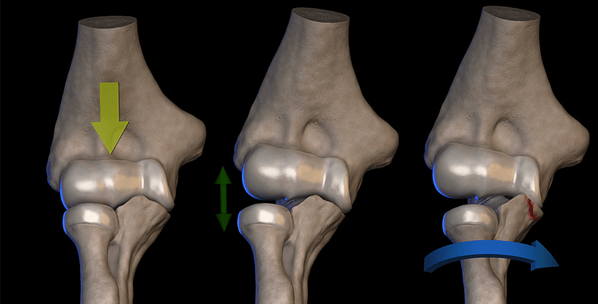
Complex instability denotes the presence of a fracture associated with a dislocation. Elbow fracture-dislocations are complex traumatic injuries of the elbow with breaks in the bones, tears of ligaments and tendons, and elbow instability.
The elbow is the second most commonly dislocated large joint.
Elbow stabilisers
⦁ Elbow stability is conferred by bony anatomy and ligaments
Primary stabilisers
⦁ The Bony architecture
⦁ The anterior band of the medial collateral ligament
⦁ The lateral ulnar collateral ligament (LUCL)
Secondary stabilisers
These include the radial head and dynamic constraints such as the muscles of the forearm.
Mechanism of injury
⦁ Most elbow dislocations occur with a fall on an outstretched arm.
⦁ Simple elbow dislocations are classified by the direction of displacement of the forearm bones about the arm, with posterolateral dislocation the most common.
⦁ Less common variants include anterior, medial, or lateral dislocations.
Types of Instability
Acute – a traumatic episode disrupts one or more of the elements contributing to stability (bones, ligaments, muscles) by placing abnormal forces on to a normal elbow.
Recurrent – as a result of a previous injury one or more of the stabilising elements is deficient. Normal forces applied to the abnormal elbow can result in repeated dislocation or subluxation of the elbow.
Simple – A dislocation with no fractures but the soft tissues (ligaments, capsule and/or muscle origins) are disrupted.
Fracture Dislocation – dislocation of the elbow with a fracture of one or more of the bones of the elbow (humerus, radius or ulna).
Considerations for Treatment
The primary aim of management for elbow instability is to restore and maintain normal articular alignment. Surgery may be required to repair anatomical structures to permit active mobilisation of the elbow within a week of the injury if possible. The general principle is to restore at least three of four structures (lateral ligament complex, radial head, coronoid process and medial collateral ligament). If the distal humerus is damaged then restoration of two out of the three following elements; medial trochlea ridge, capitellum and/or lateral trochlea ridge.
SURGICAL MANAGEMENT
Indications
⦁ Surgical management is indicated in unstable elbows, even when placed in flexion (more than 30 degrees) and pronation, elbows that recurrently subluxate or dislocate during the treatment protocol, or those with associated fractures (“complex” instability).
⦁ Management of simple dislocation requires repair or reconstruction of the ligamentous structures leading to instability. By definition, simple dislocation occurs without fracture.
⦁ An algorithmic approach to ligament repair is used to stabilise the elbow. Lateral ligament complex insufficiency is the primary lesion with simple dislocations and is therefore addressed first.
⦁ The Lateral ligament complex usually avulses from the humerus and can be repaired in the acute setting.
⦁ Repair may be performed via bone tunnels in the humerus or with suture anchors, depending on the surgeon’s preference. Prof Imam has a low threshold of augmenting these repairs with soft tissue, including tendon/ligament or synthetic ligaments. Reconstruction of the Lateral ligament complex is rarely required in acute management but is needed in chronic instability. Reconstruction is performed with one of your tendons that we can sacrifice, a similar tendon from a cadaver or a synthetic construct.
⦁ Repair or reconstruction of the Lateral ligament complex typically confers stability.
⦁ Persistent instability after Lateral ligament complex repair is rare and is more commonly observed with fracture-dislocations or chronic instability.
⦁ If persistent instability exists, the medial ligamentous complex is repaired or reconstructed.
⦁ A hinged external fixator may be placed to protect the repair.
Preoperative Planning
⦁ Clinical examination is crucial
⦁ Scans in the form of plain radiographs, CT scans and MRI scans can be done. Planning should include preparing for the possibility of reconstruction of the Lateral ligament complex, which might require reconstruction or augmentation.
⦁ A hinged external fixator should be available in the rare case that the elbow remains unstable after ligamentous repair or reconstruction.
Rehabilitation
Active elbow mobilisation should commence as soon as possible after a dislocation or fracture-dislocation, preferably by day five post-injury where possible. Stability is improved by performing exercises while lying the patient on their back with the should be flexed (brought forward) to 90 degrees.
In this position, gravity helps to maintain the congruency of the joint. Ice can help control swelling. Hinged elbow braces are not usually required and should be used only with caution under the supervision of a surgeon and physiotherapist.
A surgeon may apply an external fixator (frame) to the elbow to maintain stability in some circumstances. Casts and splints are not usually employed for initial immobilisation while awaiting stabilisation and should not be worn for more than three weeks.
SUMMARY
⦁ Elbow Dislocations are common elbow injuries which can be characterised as simple or complex depending on associated injury to nearby structures.
⦁ Diagnosis can be made with plain radiographs. CT studies can be helpful to evaluate for loose bodies or surgical planning.
⦁ Treatment is closed reduction followed by a short period of immobilisation for stable, simple elbow dislocations. Surgical management is indicated for complex elbow dislocations associated with fractures or persistent instability.


