


Absolute contraindications include general orthopedic issues, such as active infection (local or systemic), inability to undergo general anesthesia, paralysis of the arm, and inadequate postoperative soft-tissue coverage.
Relative contraindications include young age, because younger patients tend to put high-demand stress on the elbow joint by participating in sports and other activities, and diabetes and smoking, because either of these conditions might compromise wound healing. In addition, walking with crutches during rehabilitation may be problematic for patients who have had elbow arthroplasty.
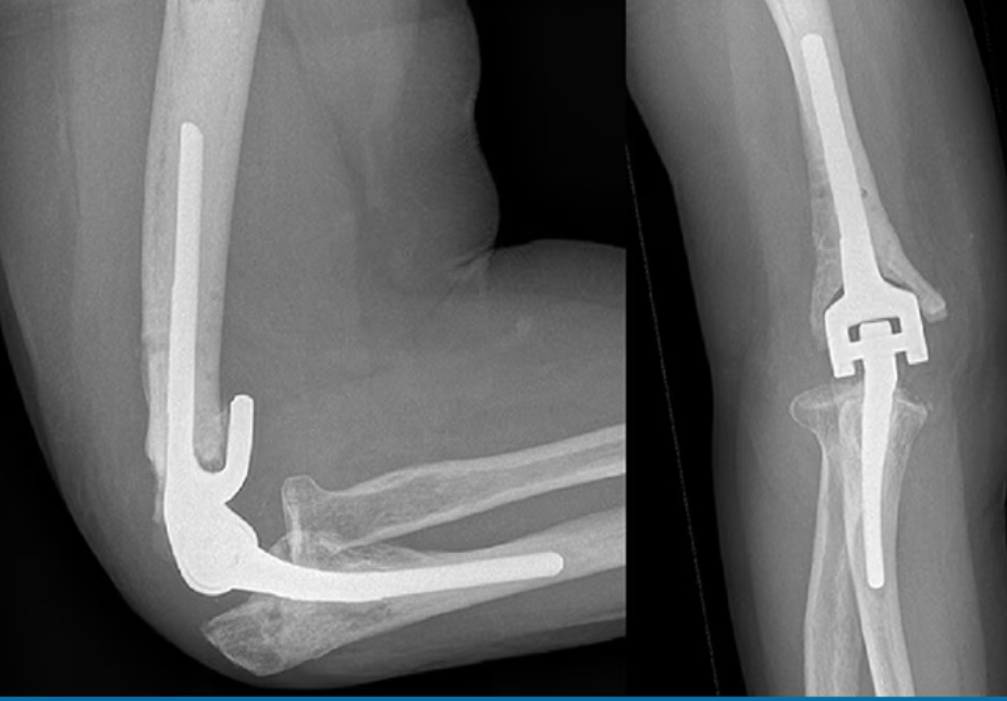
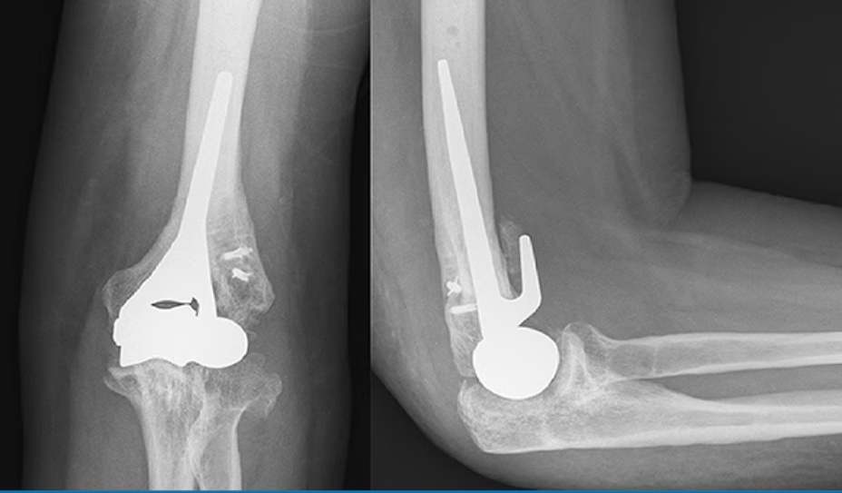
Unlinked implants have no physical connection between the humeral and ulnar components. They rely on bearing surface architecture as well as soft-tissue integrity for elbow stability.
The inherent stability of the linked designs may result in higher forces being transferred across the implant/cement and cement/bone interfaces. This is why modern designs use a “sloppy” hinge, having 7°-10°0 varus/valgus inherent laxity at the hinge section, with a minimal motion-bearing contact area; this aims to maintain intrinsic stability without the risk for early loosening.
The fixation technique may be cemented or uncemented, with a bone-ingrowth coating. Cemented designs have the advantage of instant fixation, which might be favorable in the linked designs regarding the previously mentioned pulling-out forces. However, a long cement mantle may result in elaborate surgery if revision is necessary.
Considerations for Periprocedural Care
Regarding the affected joint and patient symptoms, pain relief and/or enhancement of range of motion should be pursued, depending on the patient’s expectations and wishes. It is also necessary to inform patients that even though a joint is replaced, it can never reach the level of a healthy native joint. Therefore, activities should be adapted, and high-impact forces avoided, to decrease the chance of the implant loosening or of periprosthetic fractures. For example, patients with a total elbow arthroplasty are advised not to lift more than 5 kg at once, although this is based on empirical experience; no trials on use and the impact of use on implant survival have been published.
Postoperative short-term follow-up is important for checking on wound healing and preventing acute infections. In a 6- to 8-week period of functional rehabilitation, patients generally have fair success at pursuing activities of daily living. Long-term follow-up is important to monitor chronic problems, such as implant loosening, and systemic disease, such as rheumatoid arthritis. Especially in unlinked arthroplasties, polyethylene bushing or inlay wear and metal wear of the humeral and/or ulnar prosthesis pose a problem, because the articulating surfaces are larger than in linked designs; unlinked models have a large articulating area and not only a small hinge surface. As such, taking a patient history, physically examining the patient’s elbow for range of motion and stability, and having the patient complete a standardized questionnaire on elbow function using the patient-reported outcome measures are now routine care.
Triceps-On and Triceps-Off Techniques
To achieve good intra-articular exposure, two approach techniques can be distinguished: triceps-on and triceps-off. The triceps-on technique without olecranon osteotomy leaves the patient with a larger range of motion postoperatively, with no additional complications, even though the surgical exposure of the operative field is technically more challenging.[14]
The triceps-on Bryan-Morrey approach is performed by making a window medial to the distal triceps tendon.[15] A variant to this approach uses windows on both sides of the triceps.[14] Another triceps-sparing approach is to perform a chevron osteotomy and to reattach the osseous insertion of the triceps afterwards.
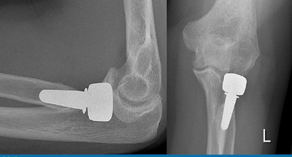
In radial head arthroplasty, it is necessary only for the humeroradial articulation to be exposed. Three usual approaches can be distinguished, with many variations possible, depending on the individual need.
The first is the posterolateral or Kocher approach, as described by Theodor Kocher in 1892.
This approach uses the interval between the anconeus and muscles, which leads to the lateral side of the radial head and proximal radius. To achieve a wider exposure, the extensor carpi ulnaris at the point of origin on the humerus or the insertion of the anconeus muscle where it inserts distally on the posterior surface of the ulna can be dissected. During this approach, the lateral collateral ligament is at risk to be damaged or lifted off of the ulna because of the subperiostal dissection.
The second approach, the anterolateral or Kaplan approach, uses the interval between the extensor carpi ulnaris and extensor digitorum communis anterior to the Kocher approach.
During this approach, the posterior interosseous nerve (PIN) shifts during pronation and supination. Therefore, to turn the PIN anteriorly, this approach is performed with the forearm in pronation.
Short- and Long-Term Complications
The most serious short-term complications are neurovascular injury, wound healing problems, infection, and dislocation.Good wound hygiene lowers the superficial infection rate, and sterile surgery, combined with antibiotic-loaded cement, decreases the rate of deep periprosthetic infections.
In the long term, loosening of the implant on either an septic or aseptic basis is the main cause of device failure.[11] In radial head replacement and hemiarthroplasty, the opposing articular bone quality must be evaluated to monitor erosion.
Implant survival rates are implant-specific, yet several trends are distinguishable. Total elbow arthroplasties currently have a mean 10-year survival rate of approximately 90%, which is an increase over previous decades. Radial head prostheses have good long-term results, but large implant registry data are lacking.
Conclusion
Especially in the past four decades, significant advances have been made in elbow arthroplasty. However, owing to the relatively small numbers of patients requiring elbow arthroplasty, regardless of type, effective treatment modalities require ongoing research. To ensure optimal patient care, elbow problems are best treated in orthopedic clinics where the surgeons are expert in performing arthroplasties.